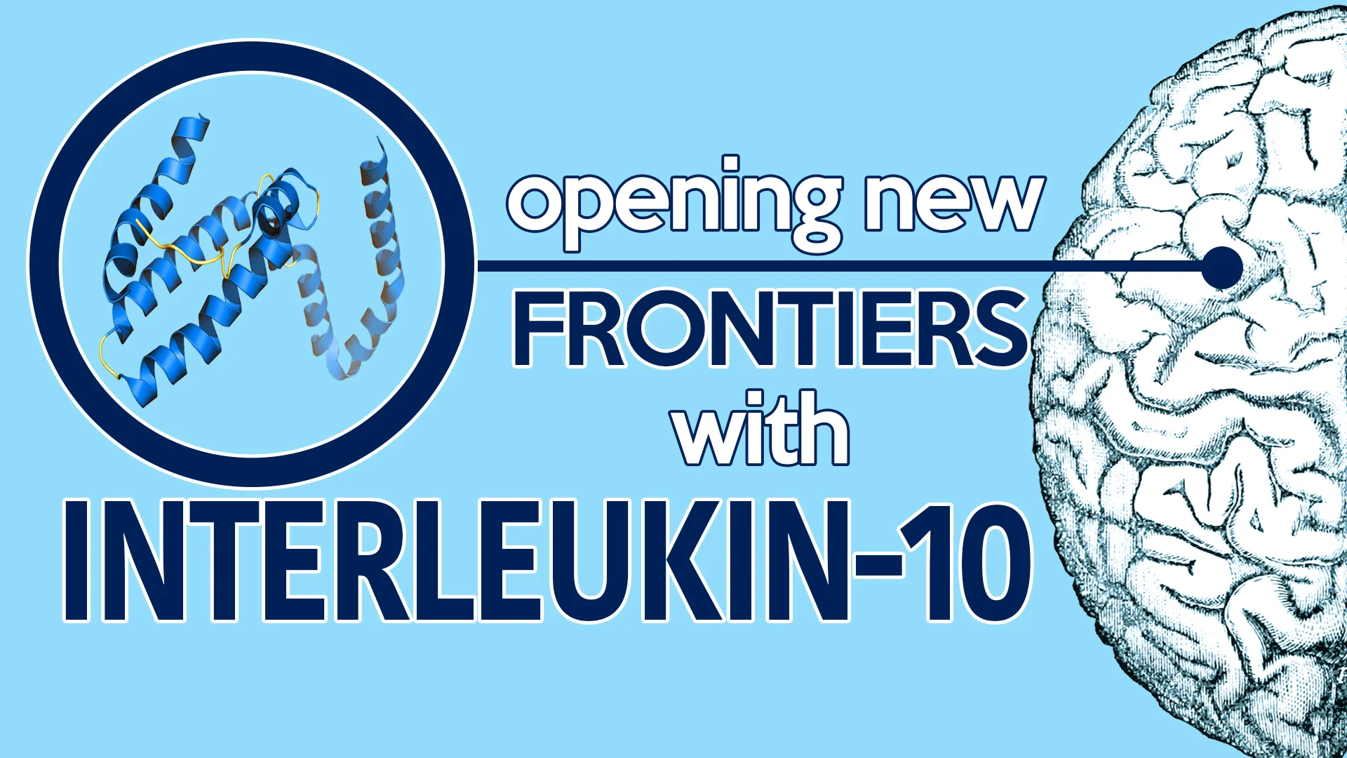
Interleukin-10 Opens New Frontiers in Alzheimer’s Research
by Joy Cui
Oliver Sacks, an American-British neurologist and lecturer at the New York University School of Medicine, once summed up the human brain perfectly: “We, as human beings, are landed with memory systems that have fallibilities, frailties, and imperfections—but also great flexibility and creativity.”
Housing close to 100 billion neurons, the brain is a powerful organ that is constantly working to coordinate communication amongst itself, the body, and the outside environment. When this expansive network of neurons within us fails to function properly, we struggle to respond to stimuli in our surroundings and carry out even simple movements. The brain likewise experiences a decline in its ability to recall memories, think and initiate social interactions.
Alzheimer’s Disease (AD) is a debilitating neurodegenerative illness that results in this very phenomenon. Affecting over five million Americans, AD is the number one form of dementia and the sixth leading cause of death in the United States. It is classically characterized by memory loss, difficulty completing everyday tasks, poor spatial judgment and various other progressive symptoms. The origin of these developments is linked to the buildup of misfolded proteins—beta-amyloid and neurofibrillary tangles—in the brain.
In contrast to healthy brains, the cerebral matter of Alzheimer’s patients cannot break down the dysfunctional beta-amyloid, which eventually forms hard, insoluble plaques. Neurofibrillary tangles, on the other hand, are composed of tau, which are microtubule proteins that have twisted fibers and cause similar manifestations. As plaques and tangles spread across the brain, intercellular signaling and delivery of nutrients are increasingly interrupted. As a result, neurons are unable to effectively communicate, initiate body movement, understand spatial relationships and put memories together, hampering some of the most core functions that shape a person’s daily life.
Traditionally, non-steroidal anti-inflammatory drugs (NSAIDs) have been hailed as the primary medical treatment for AD. These drugs are thought to work by reducing inflammation, or swelling, due to beta-amyloid plaques and neurofibrillary tangles in the brain. When inflammation exists, microglia—specialized neurons that act as a form of immune defense—move to the site. Upon arrival, they start clearing the cellular debris and attack infectious substances through phagocytosis, a cellular process that involves engulfing nearby substances. Through this process, the outer membrane of the microglia wraps around their targets to “engulf” them, forming an internal vesicle called a phagosome, which can then be digested using enzymes. Alternatively, microglia can also release cytokines, which are proteins that are able to influence the behavior of neighboring cells. This allows microglia to serve as an alert mechanism in order to contain the spread of inflammation.
Unfortunately, NSAIDs have produced mixed results in AD patients. Their exact relationship with microglia and plaques remains unclear due to evidence that they fail to definitively improve patients’ conditions.
Over the course of research in this area, activated microglia have been consistently spotted near deposits of plaque. This observation suggests that microglia-induced inflammatory responses facilitate neurodegeneration, where the neuronal damage is caused by cytokines and various substances released by microglia. While a large number of patients on NSAIDs do experience some relief, cytokines generated by microglia have been linked to triggering additional production of amyloid precursor protein, one source of beta-amyloid plaque production. Due to this possibility, NSAIDs could potentially lead to even more buildup of beta-amyloid plaque.
In recent years, some researchers have begun challenging NSAIDs as there has been increasingly more evidence that beta-amyloid plaques can be removed by blocking interleukin-10 (IL-10), one of many anti-inflammatory cytokines. Studies conducted by the University of Florida, Italy’s Università di Palermo and the University of South California’s (USC) Keck School of Medicine have all produced similar results that support the correlation between diminished expression of IL-10 and clearing of beta-amyloid plaques.
In order to analyze the effects of removing IL-10 from their study subjects, USC researchers knocked out the gene that expresses IL-10 in mice to produce mice deficient in the interleukin. Strikingly, microglia activation increased up to 266 percent in IL-10 deficient mice.
It may initially appear that blocking IL-10 would simply increase the amount of cerebral plaque. However, what USC researchers found upholds previous research findings: when IL-10 is blocked, a different, yet beneficial immune response is triggered.
When IL-10 is blocked, phagocytosis is promoted by microglia, which proceeds to engulf and digest the plaque. For instance, the researchers noted that microglia involved in plaque digestion contained 60 percent more beta-amyloid in the cortex and 62 percent more beta-amyloid in the hippocampus of mice with IL-10 blocked! These statistics imply that microglia were activated to favor phagocytosis over the release of potentially harmful cytokines. Overall, researchers concluded that there was 60 percent more beta amyloid uptake by microglia.
Currently, researchers know that microglia are less capable of clearing beta-amyloid as AD progresses. Nonetheless, the USC group also revealed that their AD-afflicted, IL-10 deficient mice behaved and scored more like mice without Alzheimer's in learning and memory tasks. Therefore, the USC group is putting forth the idea that microglia can have their phagocytic ability restored as long as IL-10 is absent. Unfortunately, the researchers did not determine whether the release of cytokines was impacted by the enhanced phagocytic role of microglia when IL-10 is blocked.
As highlighted by Terrence Town, senior author of the study, “Alzheimer's disease is the public health crisis of our time.” This astounding study not only questions the traditional idea that anti-inflammation responses should be promoted in brains that contain beta-amyloid plaque, but also provides a novel alternative pathway that revolutionizes the medical, pharmaceutical and research fields.
To Town, this study shows that the “immune response to wipe away toxic plaques from the brain may bring new hope for a safe and effective treatment for this devastating illness of the mind." While the research studies conducted in animal models do not offer a definite conclusion that similar results will be replicated if IL-10 is blocked in humans, it does offer a strong hope. Ultimately, the road ahead is long and risky, but the potential benefits are incredible and enticing.
